Your cart is currently empty!
Visual Field Scans
A Visual Field Scan is a subjective test that shows what a patient can see in their central vision field. At Arkansas Acupuncture, we track our patients’ progress by taking visual field scans before and throughout their treatment.
Before treatment, the scans help to determine the severity of visual symptoms. Throughout the treatment, the scans show how effective we are at treating vision problems.
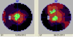
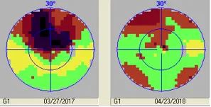
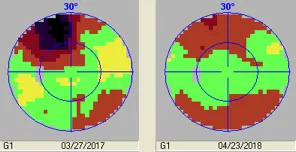
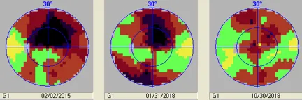
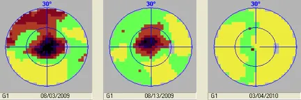
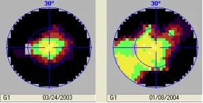
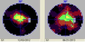
The scans below, from current and former patients, show the progression of improvement over time. In each group, the scan on the left represents the baseline, or the first scan taken at the Arkansas Acupuncture Center. The scan on the right is the most current, and the scan in the middle shows the last scan before the current scan. As you can see, there is the potential for definite improvement in vision over time.
Macular Degeneration Scans
Age-Related Macular Degeneration (AMD) is a disorder caused by the build-up of amyloid beta plaques in the back of the eye. Patiends with Age-Related Macular Degeneration will have deterioration of the cells in the macula causing a small blurry spot that, over time, can enlarge and leave the patient with only peripheral vision.
The macula is in the center, the area where we want the most vision improvement since it allows for detailed vision. The entire scan represents 30 degrees of vision; the visual field. Beyond that area our vision is mainly for movement, night vision and some color.
The scans below show how the dark, low-vision areas in the earliest scans become lighter in later scans, showing greater vision.
- Yellow = normal vision
- Green = minimal loss- detailed vision
- Red = poor vision
- Black = no vision
The changes are, as you can see, striking.

86-year-old female, left eye

86-year-old female, right eye
82-year-old male, right eye
82-year-old male, left eye
78-year-old male, right eye
77-year-old female, right eye

77-year-old female, left eye
83-year-old male, right eye
83-year-old male, left eye
Retinitis Pigmentosa Scans
Retinitis Pigmentosa (RP) is an inherited set of diseases that cause the photoreceptor cells of the retina to deteriorate. Many times patients with RP will lose their peripheral vision first. RP will progress until there is only central vision, causing tunnel vision. Over time the cells at the center of retina will be destroyed and the patient will become totally blind or have only small areas of low vision.
The scans below show how the darker areas of poor vision grow smaller over time, and the light areas of better vision increase.

22-year-old female, right eye
22-year-old female, left eye

10-year-old female, right eye

10-year-old female, left eye
16-year-old male, right eye